MODULE 1: Anatomy of the Respiratory System
INTRODUCTION
All life processes are dependent on a source of energy. In the case of man, this energy is generated from the basic components of our food supply, primarily through metabolic processes which consume oxygen and produce carbon dioxide as a waste byproduct. This is the process called respiration*. The anatomy of the respiratory system permits the body to both obtain the necessary supply of oxygen from the external environment and to eliminate excess carbon dioxide from the body.
Module Structure
This module contains only one section:
SECTION 1: Anatomy of the Respiratory System
SECTION 1: Anatomy of the Respiratory System
Introduction
In humans and animals, most energy is derived from chemical reactions involving oxygen. Cells must also be able to eliminate carbon dioxide, which is the major end product of oxidative metabolism. The circulatory and respiratory systems share the responsibility for supplying the body with oxygen and disposing of carbon dioxide. This section will focus on how the anatomical structures of the respiratory system are involved in gas exchange between the blood and external environment.
Learning Objectives
After reading this section you should be able to:
-
-
- State the function of the respiratory system.
- List the components of the upper and lower airways from the nasal cavity to the alveoli of the lungs.
- Name the structures comprising the mechanical support system.
-
General Anatomy of the Respiratory System
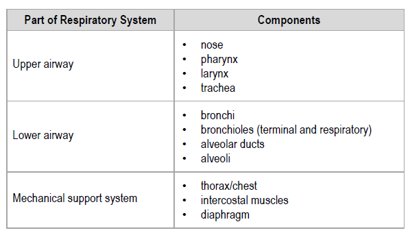
The anatomical structure of the respiratory system is divided into three subsystems:
-
-
- the upper airway
- the lower airway
- mechanical support system
-
SECTION 1A: The Upper Airway
Components
The components of the upper airway include:
-
-
- nose
- pharynx*
- larynx*
- trachea*
-
The structures of the upper airway act as passageways for the air as it moves towards the alveoli (singular alveolus*) of the lungs where gas exchange occurs.
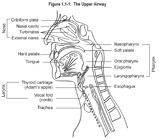
Nose
The nose consists of the external nares (nostrils) and the nasal cavity, which lies above the roof of the mouth.
The nasal cavity is actually made up of two cavities separated by a cartilaginous bony septum. In each cavity, turbinates (conchae) or bony projections extend from the sides of the cavity. The turbinates are covered with a thick mucous membrane containing epithelial cells that secrete mucus* onto the membrane surface.
The paranasal sinuses* are cavities within the facial skeleton that are lined by cilia*. The nasal cavity also contains ciliated cells.
Pharynx
The pharynx (throat) is a common channel for both the respiratory and digestive tracts. It begins at the base of the skull and extends down to the level of the esophagus*. Anatomically, it is subdivided into:
-
-
- nasopharynx* — lies behind the nasal cavity
- oropharynx* — lies behind the oral cavity
- laryngopharynx* — lies behind the larynx
-
Larynx
The larynx, commonly referred to as the “voice box”, connects the pharynx and the trachea. It extends from the epiglottis* to a point slightly below the “Adam’s apple” (thyroid cartilage). The epiglottis is a flap-like structure at the entrance to the larynx which closes during swallowing so that food goes into the esophagus. It remains open during breathing.
Within the larynx lie the vocal cords. The flow of air past the vocal cords causes them to vibrate allowing us to speak. The slit-like passageway between the vocal cords is the glottis*.
Trachea
The larynx opens into the trachea or “windpipe”. The trachea is about twelve to fourteen centimetres long. It divides into two bronchi*, the left and right main stem bronchi. At birth, the inner diameter of the trachea is only about 3 mm, reaching a diameter of about 16 mm in the adult.
The trachea is composed, anteriorly and medially, of rigid horseshoe shaped cartilaginous rings. Posteriorly, the trachea is composed of muscle and other tissues. The trachea has only a limited capacity to dilate or constrict.
The trachea is lined by a mucous membrane containing mucus-secreting cells and ciliated epithelium. The mucociliary escalator* plays an important role in clearing the airway of smaller foreign substances that gain entrance to the upper and lower airways.
SECTION 1B: The Lower Airway
Components
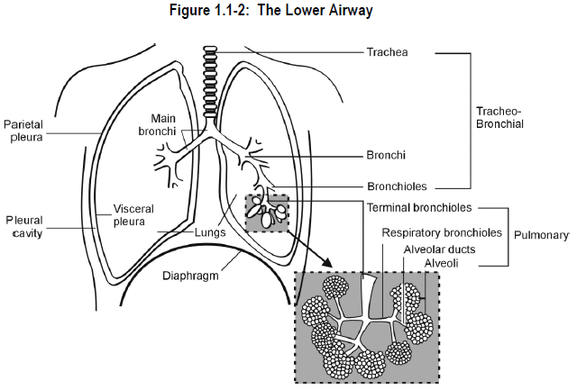
In descending order of size, the components of the lower airway include:
-
-
- bronchi
- bronchioles
- terminal bronchioles*
- respiratory bronchioles*
- alveolar ducts
- alveoli
-
All of these structures are located within the right and left lungs.
Lungs
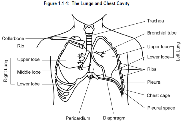
Despite their relatively large size, the lungs weigh only about 2.5 pounds. Healthy lungs are soft and spongy in appearance. The right lung is comprised of three lobes and is usually larger than the left lung, as it is responsible for about 55% of total ventilation*. The left lung has two lobes and accounts for the remaining 45% of total ventilation.
Each lung is surrounded by a completely closed sac called the pleural sac which consists of a shiny, thin sheet of cells called the pleura. The surface of each lung is covered with a visceral serosa called the visceral pleura and the walls of the thoracic cavity are lined by the parietal pleura.
The space between the two pleural sacs is called the pleural cavity. The pleural cavity contains a thin layer of fluid called the pleural fluid. This fluid lubricates the outer surface of the lungs. Pressure changes in the pleural fluid cause the lung surface and thoracic wall to move in and out together during breathing.
Bronchi
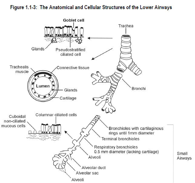
The structures of the lower airway serve to conduct air from the gas transporting system of the upper airways into the gas exchange areas of the lower airways. The anatomical and cellular structures of the lower airways are shown in Figure 1.1-3.
The left and right main stem bronchi split off from the base of the trachea and continue to branch off into various sections of each lung.
As the bronchi become narrower, the cartilaginous rings present in the trachea diminish and are replaced by an increase in smooth muscle* which is capable of contracting or relaxing in response to nervous stimulation or various chemical substances.
The lining of the bronchi also becomes less ciliated and the surface epithelium becomes thinner.
Bronchioles
When the bronchi no longer have cartilage, usually when they reach an inner diameter of less than 1 mm, they are called bronchioles. This constitutes the level at which the “small airways” are said to begin. The smooth muscle in their walls is circumferential. When it contracts, it makes the air tube smaller.
1. Terminal bronchioles
Just prior to the area where gas exchange occurs, the bronchioles reach an inner diameter of about 0.5 mm. These are called terminal bronchioles. They have few cilia in their epithelial lining and have smooth muscle as the major component of their wall. They do not have alveoli in their wall.
2. Respiratory bronchioles
The terminal bronchioles further subdivide into small bronchioles termed respiratory bronchioles which connect the terminal bronchioles to the alveolar ducts. Respiratory bronchioles themselves have alveoli within their wall (hence they are called “respiratory”). The alveolar ducts, in turn, connect with the gas exchange units of the lung known as alveoli. At the respiratory bronchiole level, the units lack smooth muscle and are simply epithelial tubes. Their lumens are held open by the subatmospheric pressure of the thoracic cavity.
Alveoli
The alveoli, or air sacs, lie at the end point of the bronchiole tree. There are millions of clustered alveoli, which resemble bunches of grapes. The alveoli maximize the surface area available for gas exchange, which has been estimated to be between 40 to 100 square meters in the adult. The alveoli are lined by a single thin layer of epithelial cells. The external surface of the alveoli is covered in “cobwebs” of pulmonary capillaries through which gas is exchanged. Gas exchange is explained more fully in Module 2.
SECTION 1C: Mechanical Support System
Structures
The mechanical support structures include the diaphragm* and chest wall. They play a vital role in controlling pressure within the chest (thoracic cavity). Changes in intrathoracic pressure are responsible for moving air in and out of the lungs.
Thorax/Chest
The lungs and the heart are situated in the thorax, the compartment of the body that lies between the neck and abdomen. The terms “thorax” and “chest” are used interchangeably. The chest wall is formed by the spinal column, the ribs, the sternum (breast bone) and the intercostal muscles that lie between the ribs. The thoracic wall also contains large amounts of elastic connective tissue.
Diaphragm
The diaphragm, as shown in Figure 1.1-4, is a large sheet of muscle that separates the abdomen from the thoracic cavity. The diaphragm is the major muscle of inspiration*. It contracts and descends on inspiration. This stretches the lungs, which then passively return to rest during expiration*.
The diaphragm is the most important muscle for breathing.
If the diaphragm should fail due to obstructive pulmonary disease or neuromuscular disease, the accessory muscles of breathing are called upon at great energy cost to the patient.
SUMMARY — SECTION 1: Anatomy of the Respiratory System
The respiratory system, in conjunction with the circulatory system, supplies the cells of the body with oxygen and disposes of carbon dioxide.
The respiratory system is divided into three subsystems:
-
-
- the upper airway — passageway for air
- the lower airway — gas exchange
- the mechanical support system — pressure control within the thoracic cavity
-
PROGRESS CHECK — SECTION 1: Anatomy of the Respiratory System
1.
What is the principal function of the respiratory system?
____________________________________
____________________________________
2.
List the three distinct components of the respiratory system.
1. ___________________________________
2. ___________________________________
3. ___________________________________
3.
Listed below are 10 components through which air passes from the time it is inhaled to the time it is released from the body. Number the process from 1-10 with #1 starting the process.
______ alveolar ducts
______ nares
______ pharynx (including naso-, oro- and laryngopharynx)
______ terminal bronchioles
______ alveolar sacs
______ lungs via trachea (into 2 bronchi)
______ larynx
______ nasal passages
______ respiratory bronchioles
______ nasal cavity
4.
List the components of the mechanical support system.
___________________________________
___________________________________
___________________________________
PROGRESS CHECK ANSWERS — SECTION 1: Anatomy of the Respiratory System
1.
The principal function of the respiratory system is to supply the cells of the body with oxygen and to dispose of the carbon dioxide.
2.
Components of the respiratory system:
1. upper airway
2. lower airway
3. mechanical support system
3.
Components through which air passes:
1. nares
2. nasal cavity
3. nasal passages
4. pharynx (including naso-, oro- and laryngopharynx)
5. larynx
6. lungs via trachea (into 2 bronchi)
7. terminal bronchioles
8. respiratory bronchioles
9. alveolar ducts
10. alveolar sacs
4.
Components of the mechanical support system:
thorax/chest wall
intercostal muscles
diaphragm
MODULE 1 Test
1.
All of the following structures are considered to be part of the upper airway EXCEPT:
a) alveoli
b) larynx
c) trachea
d) pharynx
2.
Which of the following is a common channel for both the respiratory and digestive systems?
a) trachea
b) larynx
c) nasal cavity
d) pharynx
3.
What structure guards the entrance to the larynx, closing during swallowing to prevent entry of food and other substances into the lungs?
a) alveolus
b) epiglottis
c) trachea
d) vocal cords
4.
What is the correct order of DESCENDING SIZE of the following airways?
a) bronchioles, bronchi, alveolar ducts, terminal bronchioles, respiratory bronchioles
b) bronchi, respiratory bronchioles, terminal bronchioles, alveolar ducts, bronchioles
c) bronchi, bronchioles, terminal bronchioles, respiratory bronchioles, alveolar ducts
d) alveolar ducts, bronchi, bronchioles, respiratory bronchioles, terminal bronchioles
5.
All of the following statements are true EXCEPT:
a) alveoli are the gas-exchange units of the lung
b) the alveoli are lined by a single thin layer of epithelial cells
c) the alveoli maximize the surface area for gas exchange
d) capillaries empty blood into the alveoli where gas exchange occurs
6.
Which of the following muscles separates the abdomen from the thoracic cavity?
a) intercostal muscle
b) diaphragm
c) smooth muscle
d) trachealis muscle
MODULE 1 Test Answers
1.
a) alveoli
2.
d) pharynx
3.
b) epiglottis
4.
c) bronchi, bronchioles, terminal bronchioles, respiratory bronchioles, alveolar ducts
5.
d) capillaries empty blood into the alveoli where gas exchange occurs
6.
b) diaphragm
