Module 1: Anatomy and Physiology of the Skin
Introduction
In this module, we discuss the structure and various functions of the skin and its major appendages.
Module Structure
This module is divided into two sections:
Section 1: Structure of the Skin
Section 2: Functions of the Skin
Learning Objectives
After reading this section, you should be able to:
- List the major components of the skin.
- Describe the structure of the epidermis, dermis and subcutaneous layer and state the function of each.
- List the major appendages of the skin and state the function of each.
Components of the Skin
Skin consists of three layers:
1) Epidermis
The epidermis is the surface layer of the skin. It is thin and tough and provides protection for the layer below.
2) Dermis
The dermis contains structures that help control the body’s temperature and sense its surroundings. It also is responsible for lubricating the skin.
3) Subcutaneous layer
The epidermis and dermis comprise the skin. The subcutaneous layer connects the skin to the surface muscles.
Epidermis
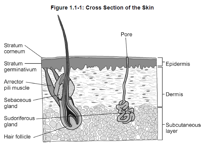
The epidermis is the outer, thinner layer of the skin and is made up of several layers. These include the:
-
-
- stratum corneum
- stratum germinativum
-
1) Stratum corneum
This outermost layer of the skin contains only epithelial cells and no blood vessels. The surface cells are dead cells that are constantly being sloughed off due to wear and tear on the skin.
The stratum corneum is also referred to as the horny layer because the cells are very flat and horny in appearance.
2) Stratum germinativum
The stratum germinativum contains cells that are constantly undergoing mitosis* to produce new daughter cells. This layer is also called the growing layer. The cells receive nourishment from the dermis.
As the daughter cells are pushed up towards the stratum corneum, they start to undergo changes due to lack of nourishment. The cells flatten and harden, and eventually die and are sloughed off. Large amounts of the protein keratin* are formed. Keratin hardens, thickens and protects the skin. This process is called keratinization*. Keratin is able to absorb water and helps to keep the skin hydrated.

Melanin* is the pigment that gives skin its colour. It is produced by melanocytes located at the border of the epidermis and dermis. The melanocytes become more active on exposure to sunlight and produce melanin to help protect the dermis from ultraviolet light (“tanning effect”).
Dermis
The dermis is also called the corium. It is a layer of fibrous connective tissue that is deeper and thicker than the epidermis. It contains elastic fibres and collagen* fibres.
The dermis is well supplied with blood vessels, and nerve endings and specialized sensory receptors sensitive to touch, pressure, pain and temperature.
This layer protects the body from mechanical injury, supports the epidermal structures and provides elasticity to the skin.
The dermis is composed of both cellular and fibrous components. Fibrocytes, histiocytes and mast cells comprise the cellular components of the dermis. Fibrocytes, which are responsible for the structure of the dermis, are the most important dermal cells.
Histiocytes are mobile cells with phagocytic properties. The mast cells, which are found around blood vessels and hair follicles*, play an important role in inflammatory reactions.
The fibrous structure of the dermis is composed of collagen and elastic fibres embedded in an extrafibrillar gel made up of proteoglycans and glycoproteins. The polysaccharides in the proteoglycans contain hyaluronic acid*, a substance that attracts and retains water. In fact, the dermis contains large amounts of water and serves as a water storage site. Fibronectin, one of the glycoproteins, plays an important role in the healing of wounds.
Subcutaneous Layer
The subcutaneous layer below the dermis is composed of loose connective tissue, including adipose tissue*. Adipose tissue helps to insulate the body and aids in temperature control by minimizing both heat gain and heat loss. The adipose tissue provides for storage of energy and nutrients.
Appendages
The skin also contains appendages or accessory structures. Most of these appendages are located in the dermis and may extend into the subcutaneous layer.
1) Sudoriferous glands
Sudoriferous glands, or sweat glands, are coiled, tubelike structures located in the dermis and the subcutaneous layer.
There are two types of sudoriferous glands:
-
-
- eccrine glands
- apocrine glands
-
Eccrine glands have a duct that opens directly on the skin surface at a pore. They are located throughout the body. The secretion by these glands of varying amounts of sweat containing water and some mineral salts helps to regulate body temperature and water content of the body.
Apocrine glands are located mainly in the armpits and the groin area. These glands release their secretion into hair follicles in response to emotional stress and sexual stimulation. The secretion contains cellular material broken down by bacteria and is a cause of body odour.
2) Sebaceous glands
Sebaceous glands, or oil glands, are saclike structures with ducts that mainly open directly into the hair follicle. These glands produce an oily secretion called sebum*, which lubricates the skin and hair and prevents drying.
3) Hair
A hair is composed of dead, hardened epidermal cells. Hair contains hard keratin that does not slough off like the softer keratin covering the surface of the epidermis. Each hair develops within a follicle and new hair is formed from the hair root located at the bottom of the follicle. Each follicle has one or more sebaceous glands. Also attached to most hair follicles is an involuntary smooth muscle called the arrector pili muscle. These muscles are innervated by sympathetic nerves. Upon exposure to cold temperatures or emotion, these muscles contract causing hair to be raised and producing “goose bumps”. The muscle also presses on the sebaceous gland causing the release of sebum.
4) Nails
Nails contain hard keratin produced by cells originating in the stratum corneum. New cells are being formed continuously in the nail matrix.
Summary — Section 1: Structure of the Skin
Skin consists of three layers:
-
-
- epidermis – outer thinner layer made up of several layers, including the stratum corneum and stratum germinativum
- dermis – contains elastic fibers and collagen fibers
- subcutaneous layer – deepest layer composed of connective and adipose tissue
-
The epidermis contains melanin, which provides protection from ultraviolet rays, and keratin, which helps keep the skin hydrated. The epidermis also produces new skin cells.
The dermis protects the body from mechanical injury, supports the epidermal structures and provides elasticity to the skin.
Adipose tissue in the subcutaneous layer insulates the body, helps to regulate body temperature and stores energy and nutrients.
Skin appendages, mainly located in the dermis, include:
-
-
- sudoriferous glands – sweat glands
- sebaceous glands – oil glands, which secrete sebum
- hair
- nails
-
Progress Check — Section 1: Structure of the Skin
1.
List the function of each of the following skin components.

2.
Two of the layers of the epidermis are called the ______________________ and the ______________________.
3.
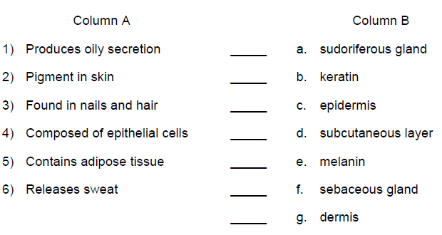
Match the description in Column A with the correct skin structure in Column B.

Progress Check Answers — Section 1: Structure of the Skin
1.

2.
stratum corneum
stratum germinativum
3.
1) f. sebaceous gland
2) e. melanin
3) b. keratin
4) c. epidermis
5) d. subcutaneous layer
6) a. sudoriferous gland
Section 2: Functions of the Skin
Learning Objectives
After reviewing this section you should be able to:
- Describe the role of the skin in protecting the body against the environment, reducing susceptibility to infections and preventing fluid loss.
- Explain how the hydrolipid film and natural moisturizing factors ensure cutaneous hydration.
- Describe the role of the skin in maintaining skin pH.
- Describe the role of the skin in maintaining body temperature.
- Describe the role of the skin in sensing the body’s environment.
- Describe the role of the skin in the synthesis of vitamin D.
- Describe the percutaneous absorption of substances through the skin.
Protection
Perhaps the most important function of the skin is protection from our environment. The skin is frequently subjected to mechanical attack and exposed to chemicals, radiation and infectious microorganisms. Under normal circumstances, the skin responds to these various stimuli with an increase or decrease in its ability to provide protection.
1) Protection against the environment
The stratum corneum provides the greatest protection against harmful substances in the environment. For example, when the skin is repeatedly exposed to mechanical attack, as when the skin of your hands is rubbed repeatedly on the handle of a shovel, it reacts by increasing the thickness of the cornified layer of the epidermis.
Protection from the sun is achieved by increasing the production of melanin. This process, called melanogenesis*, takes place within the melanocyte. During melanin synthesis, the enzyme tyrosinase reacts with tyrosine to form lipoproteins called melanosomes. Melanosomes are transformed into melanin by a series of oxidative chemical reactions.
Melanin granules are transferred to the adjacent epidermal cells to provide the skin’s normal pigmentation. There are several different kinds of melanin:
-
-
- eumelanin causes brown or black skin pigmentation
- phaomelanin gives a more reddish pigmentation
-
Melanin plays an important photoprotective role. Skin that is naturally highly pigmented (e.g., African Americans) has increased protection from the radiation effects of the sun, when compared with skin that is naturally pigmented only lightly (e.g., peoples of Celtic descent).
2) Protection from infection
Each of the following factors helps to reduce the skin’s susceptibility to infection:
-
-
- histiocytes (macrophages) in the dermis
- the oily emulsion that normally covers our skin
- normal microbial flora of the skin (normal skin flora such as Propionibacterium acnes and Staphylococcus epidermidis help to inhibit growth of pathogenic microorganisms such as Staphylococcus aureus)
- the low pH of the stratum corneum, related to the secretion of lactic acid by sweat glands, provides additional resistance to bacterial invasion
-
3) Prevention of fluid loss
Skin also provides protection through retention of body water (prevention of fluid loss) and serves as a reservoir for water. Approximately 600 mL of body water can be lost to the environment each day, even in the absence of sweating; losses during sweating can be as great as 4000 mL per hour.
Although body water is lost to the environment through perspiration, the majority of body water is retained by the protective skin barrier. This essential function is noticeably absent in patients with damaged skin surfaces. Individuals with significant areas of burned skin may lose as much as 30 litres of water per day to the environment as a result of damaged integument.
Hydration
Cutaneous hydration is an important regulatory function of the skin. Under normal conditions the stratum corneum contains 10 to 20% water. This percentage may vary according to an individual’s age and the relative humidity. If the water content drops below 10%. the skin becomes dry and cracked. When skin is dry and cracked, its protective ability is reduced considerably. Skin hydration is ensured by two mechanisms:
-
-
- the hydrolipid film
- natural moisturizing factors
-
1) Hydrolipid film
The hydrolipid film coats the surface of the skin. It is composed of fats secreted by the sebaceous glands, fatty acids from perspiration, and keratinocyte degradation products. When these lipids combine with water secreted by the sweat glands, a protective emulsion is formed.
2) Natural moisturizing factors
Natural moisturizing factors are substances that attract and retain water (hygroscopic substances). These substances make up approximately 15 to 20% of the stratum corneum.
Respiration
The skin takes in oxygen and gives off carbon dioxide. If the skin surface is occluded for a prolonged period, carbon dioxide may accumulate on the surface of the epidermis. This would lead to an increase in the pH* (i.e., a decrease in acidity) of the skin. An increase in skin pH may cause an overgrowth of pathogenic bacteria, and subsequent infection.
Thermoregulation
Normal body temperature is approximately 37 degrees Celsius. The skin, in conjunction with several neurologic and endocrine systems, helps to ensure that body temperature remains relatively constant. Skin tissue reacts to cold with vasoconstriction and increased sebaceous gland secretion, while heat stress causes vasodilation and increased sweat gland secretion.
In order for sweat to exert its cooling effect, the sweat must evaporate. If the environment is humid, evaporation of sweat and the process of body cooling are inhibited.
If, however, an individual is acclimatized to the heat and humidity, the body temperature does not rise to the same extent. This is because sweating begins sooner, and the volume of sweat produced is greater than that in a person who is not acclimatized to those environmental conditions.
Sensation
The skin is an important component of the sense of touch. Skin is extensively innervated, and is capable of registering a variety of sensations from the environment. Receptors may be either isolated or appended to hairs or sweat glands. The majority of sensory stimuli are received from free nerve endings. Sensations identified by these free nerve endings include pain and itching.
Vitamin D Synthesis
Vitamin D3 is formed when ultraviolet B (UVB) radiation from sunlight reacts with 7-dehydrocholesterol (found primarily in the epidermis).
Most Canadians receive their vitamin D through dietary sources, because of limited skin exposure to sunlight (associated with clothing for the Canadian climate plus decreased outdoor living).
Vitamin D3 is further metabolized in the body to hormonal substances that augment dietary calcium absorption. People who lack vitamin D may develop deficient mineralization of their bones (e.g., rickets* in children or osteomalacia* in adults).
Absorption
When treating skin disorders with topical drugs, absorption to the subcutaneous level is generally all that is desired. This is because the intent is to treat one or more layers of the epidermis. Drugs that penetrate the epidermis soon reach the vascular dermis, at which point the drug may be taken up by the bloodstream and distributed throughout the body. Research in dermatology has focused on making drug delivery more selective. Targeting a drug to the skin surface, epidermis, or dermis can increase the amount of drug that reaches the site of action. If drug delivery is more selective, systemic and cutaneous side effects may be reduced.
Percutaneous absorption is the process by which a substance moves from the skin surface to the systemic circulation. The skin acts as a semipermeable membrane that may be penetrated by certain substances, including medication. The primary paths for percutaneous absorption are intracellular (through the cornified cells) and intercellular (between these cells). This explains why absorption is more rapid in areas of the body where the stratum corneum is thinner or absent, such as the mucous membranes, whereas absorption is reduced where the stratum corneum is thicker (e.g., hyperkeratosis* or hyperplasia*). Although some absorption takes place along hair follicles or through sweat ducts, these pathways are generally regarded as being less significant. Absorption through these epidermal components may become substantial when the topical preparation contains a large amount of surfactant*, or if the preparation is applied to a very hairy region.
There are three basic principles of percutaneous absorption:
-
-
- neutral molecules penetrate more easily than electrically charged ions
- small molecules penetrate more easily than large ones
- molecules that are both water-soluble and fat-soluble penetrate more rapidly than either water-soluble or fat-soluble molecules
-
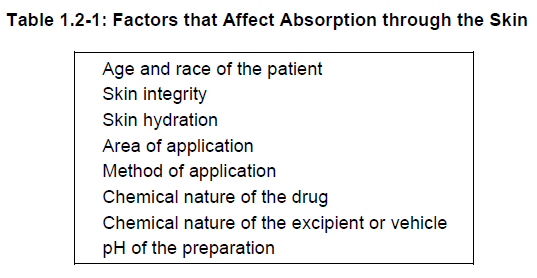
Table 1.2-1 lists the principal factors that affect absorption of substances through the skin.

Penetration of the intact drug through the skin into the bloodstream is required for transdermal drug delivery systems. One way to accomplish drug transfer across the protective skin barrier is through the use of liposomal drug carriers. Liposomes are microscopic spheres that are manufactured in layers of water (aqueous) and fat (lipid). The number and composition of these layers in a liposome varies according to the nature of the drug and its target site. Lipid soluble drugs are usually dissolved in a lipid layer, while water soluble drugs would be contained in an aqueous layer. Liposomes can also incorporate both lipid and water soluble compounds. Liposomes can act as slow-release vehicles, they can increase drug efficacy by targeting the drug to a specific cell layer, or they can localize the drug to reduce side effects. Liposomes have been used in dermatology for drugs such as corticosteroids, antifungals, retinoids, and minoxidil. The greatest use of liposomes has been in the field of cosmetics, where moisturizers contained in multilayered liposomes are being used to improve moisture penetration into the stratum corneum.
Summary — Section 2: Functions of the Skin
The skin performs many functions such as:
-
-
- protects the body from radiation, harmful substances, mechanical attack and infection
- prevents water loss and serves as a reservoir for water
- takes in oxygen and gives off carbon dioxide
- helps regulate body temperature
- provides sense of touch
- aids in formation of vitamin D
- acts as semipermeable membrane through which certain medications can be absorbed
-
Progress Check — Section 2: Functions of the Skin
1.
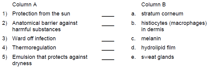
Match the description in Column A with the correct term in Column B.

2.
During melanogenesis, melanin is formed from lipoproteins called ______________________.
3.
How does the skin ensure cutaneous hydration?
________________________________
________________________________
________________________________
________________________________
4.
Why is adequate release of carbon dioxide from the skin important?
________________________________
________________________________
________________________________
________________________________
5.
How does the skin react to cold?
________________________________
________________________________
________________________________
________________________________
6.
The skin is able to form vitamin D3 upon adequate exposure to __________________ radiation.
7.
How are drugs that are applied to the skin distributed to the rest of the body?
________________________________
________________________________
________________________________
________________________________
Progress Check Answers — Section 2: Functions of the Skin
1.
1) c. melanin
2) a. stratum corneum
3) b. histiocytes (macrophages) in dermis
4) e. sweat glands
5) d. hydrolipid film
2.
melanosomes
3.
A hydrolipid film coats the surface of the skin and forms a protective emulsion. Natural moisturizing factors attract and retain water.
4.
If the skin is unable to adequately release carbon dioxide, the pH of the skin increases. This may result in excessive growth of bacteria, and potentially lead to infection.
5.
Skin tissue reacts to cold by constricting blood vessels and increasing sebaceous gland secretion.
6.
ultraviolet B
7.
Drugs penetrate the epidermis and are taken up by the bloodstream in the vascular dermis. Once they enter the systemic circulation, they are distributed to all parts of the body.
Module 1 Test
1.
The innermost layer of the epidermis is called the:
a) stratum corneum
b) stratum germinativum
2.
Keratinization is a process that involves:
a) production of melanin
b) flattening and hardening of epidermal cells
c) production of new daughter cells
d) production of sweat by glands
3.
Which of the following statements regarding the dermis is true?
a) the dermis contains elastic fibres and collagen fibres
b) hyaluronic acid in the dermis attracts and retains water
c) fibronectin in the dermis aids in wound repair
d) all of the above
4.
Another name for sweat glands is:
a) sudoriferous glands
b) sebaceous glands
5.
The substance that protects the skin from ultraviolet rays is:
a) keratin
b) melanin
c) adipose tissue
d) sebum
6.
Which of the following statements is true?
a) melanocytes are located in the subcutaneous tissue
b) melanosomes produce melanin
c) the skin reacts to cold by increasing peripheral blood flow through vasodilation
d) natural moisturizing factors prevent moisture loss from the stratum corneum by acting as an impermeable barrier to water
7.
A reduction in cutaneous respiration caused by prolonged occlusion of the skin is likely to:
a) increase the pH of the stratum corneum
b) decrease the pH of the stratum corneum
c) decrease Vitamin D synthesis
d) increase Vitamin D synthesis
8.
Vitamin D synthesis takes place in the:
a) epidermis
b) dermis
c) hypodermis
d) subcutaneous tissue
9.
Which of the following statements is true?
a) electrically charged ions have higher percutaneous absorption than neutral molecules
b) the hydrolipid film coating the skin is composed mainly of proteins
c) the skin acts as a semipermeable membrane during percutaneous absorption
d) sweat evaporates more readily in humid conditions
10.
Which of the following is a theoretical advantage of liposomal drug delivery?
a) increased efficacy
b) decreased systemic side effects
c) prolonged drug release
d) all of the above
Module 1 Test Answers
1.
b) stratum germinativum
2.
b) flattening and hardening of epidermal cells
3.
d) all of the above
4.
a) sudoriferous glands
5.
b) melanin
6.
b) melanosomes produce melanin
7.
a) increase the pH of the stratum corneum
8.
a) epidermis
9.
c) the skin acts as a semipermeable membrane during percutaneous absorption
10.
d) all of the above
