Module 1: Anatomy and Physiology of the Brain
Module Introduction
Anatomically, the nervous system is subdivided into the central nervous system and the peripheral nervous system. The central nervous system (CNS) consists of the spinal cord and the brain (see Figure 1-1).
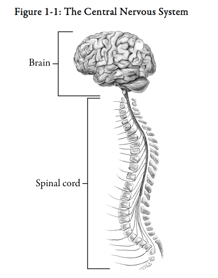
The brain processes information derived from the environment and the rest of the body, and makes decisions for behavioural strategies. The peripheral nervous system consists of nerves that fan out from the brain and spinal cord.
The peripheral nervous system relays information from body organs and the environment to the brain, either directly or via the spinal cord, and then transmits information from the CNS back to the muscles and organs.
In this module, we focus on the major anatomical and functional divisions of the brain. We discuss which areas of the brain control important body functions. We also discuss the role of nerve cells in the central nervous system in transporting important information to and from the brain.
Module Structure
This module contains two sections:
Section 1: Anatomical and Functional Organization of the Brain
Section 2: Neurotransmission
Section 1 – Anatomical and Functional Organization of the Brain
Learning Objectives
After reading this section, you should be able to:
- Identify the major anatomical and functional divisions of the brain.
- Relate specific areas of the brain to the control of specific body functions.
- Explain the importance of the blood-brain barrier.
Brain
The brain is the body’s central computer, controlling both thought and movement. The brain receives internal information from the rest of the body through sensory nerves and integrates this with external information received from the sensory organs: the eyes, ears, nose, etc. After processing this information the brain transmits instructions for action through motor nerves to the muscles and organs of the body. These responses can be voluntary or involuntary. Many body processes, such as breathing and digestion, are primarily on “automatic pilot” and do not require conscious, voluntary control. Other functions, such as movement of the limbs, are generally controlled on a conscious, voluntary basis.
There are three main structures in the brain (in ascending anatomical order):
-
-
- brainstem*
- cerebellum*
- cerebrum* or forebrain
-
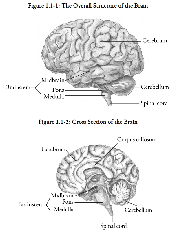
Figures 1.1-1 and 1.1-2 outline the physical relationship between these three components. We’ll discuss each of these structures in more detail.
Did you know?
The adult brain weighs about 3 pounds and contains about 10 billion nerve cells.
Brainstem
The brainstem (“primitive brain”) is the lowest section of the brain. It consists of a long stalk of nerve cells and fibres that are continuous with the spinal cord. The brainstem joins the upper spinal cord to the rest of the brain.
The brainstem has two functions:
-
-
- provides a pathway for messages from the brain to the body and the body to the brain
- connects with 10 of the 12 pairs of cranial nerves that extend from the brain and which control basic functions such as breathing, blood pressure, eye reflexes, etc.
-
The brainstem and spinal cord coordinate information between the periphery (i.e., muscles and organs) and other components of the brain. They can also function relatively independently of the brain. For instance, an animal with a brainstem and spinal cord, but no forebrain (part of the cerebrum), can stand and walk; however, these responses are aimless, not directed toward appropriate stimuli.
The brainstem consists of three main parts (in ascending anatomical order):
-
-
- medulla*
- pons*
- midbrain*
-
1) Medulla
The medulla (also called the medulla oblongata) extends from the top of the spinal cord. Within the medulla are some of the cross-roads for the nerve tracts that carry impulses to and from the brain, resulting in the right side of the body being linked up with the left side of the brain and vice versa.
The medulla also has groups of nerve cells that are involved in the following:
-
-
- taste sensation
- muscle movement in the tongue and neck
- regulation of heartbeat
- regulation of breathing
- regulation of blood pressure
- regulation of digestion
- regulation of swallowing/vomiting
-
2) Pons
The pons is a structure continuous with the medulla and anterior to the cerebellum. This structure acts like a bridge, relaying information from the medulla and cerebellum to the higher centres of the brain (cerebrum).
The pons has groups of nerve fibres that are involved in the following:
-
-
- transmission of information from the ear, face, teeth
- movement of the jaw
- adjustment of facial expression
- production of some eye movements
- co-ordination of reflex activity (e.g., urinary bladder function)
-
3) The midbrain
The area above the pons, constituting the last section of the brainstem, is referred to as the midbrain. This area contains nervous centres (e.g., the substantia nigra*) that are of great importance in motor co-ordination, serving to integrate motor signals from other parts of the brain. Several of these nervous centres function as relay stations for impulses from the eyes and ears to higher centres of the brain.
Did you know?
The brainstem is 7.5 cm (3 in) long and is about the diameter of a thumb.
Cerebellum
The cerebellum (“little brain”) is a large folded structure attached to the dorsal side of the pons. The cerebellum has an outer grey matter structure with several layers like the bark of a tree.
If the cerebellum is damaged, a variety of coordination deficits are found. In none of these deficits is there a total loss of movement, but rather a loss in the speed, the smoothness, or the strength of the movements. For example, in patients with cerebellar disease there is a tendency to sway when walking. Tremors and jerks may accompany this movement. There is difficulty in keeping eyes on targets and in speaking clearly and smoothly. These deficits suggest that the cerebellum supplies information to keep movements smooth and accurate, and to make small corrections while they are in progress. The cerebellum receives constant sensory information from all regions of the body.
Cerebrum
The cerebrum is the largest structure of the brain, comprising 85 percent of the weight of the brain. Specific areas of the cerebrum control conscious mental processes, sensations, emotions, and voluntary movements.
1) Cerebral hemispheres
The cerebrum is divided into the right and the left hemispheres by the medial longitudinal fissure. See Figure 1.1-3.
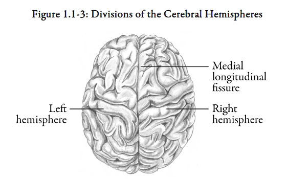
The cerebral hemispheres process information differently. The left cerebral hemisphere specializes in symbols and logic and has given humans science and technological capacity. The right cerebral hemisphere specializes in pattern and space perception and has given us art and the capacity for imagination. Beliefs also reside in this area of the brain.
These hemispheres are connected only in their lower middle portion by the corpus callosum* (shown in Figure 1.1-2), a mass of nervous fibres that enables the two sides of the brain to communicate.
2) Lobes of the cerebral hemispheres
Each cerebral hemisphere is divided into four lobes by small fissures known as sulci (singular, sulcus):
-
-
- frontal lobe
- parietal lobe
- occipital lobe
- temporal lobe
-
See Figure 1.1-4.
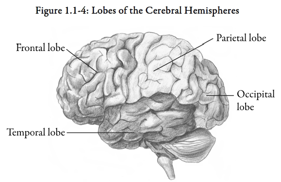
Bumps between the sulci are called gyri (singular, gyrus). Each lobe has been found to control certain brain functions.
The frontal lobe has grown especially large in humans, occupying the regions behind the forehead. This region is important for long-term plans and for complex moral judgments. These traits distinguish us from other species.
3) Cerebral cortex
The outer layer of grey matter of the cerebrum is referred to as the cerebral cortex*.
-
-
- Visual cortex
-
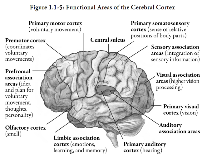
The sensory and motor areas of the cerebral cortex are shown in Figure 1.1-5. At the posterior of the brain is a large area involved in vision. The largest sub-area of the visual cortex, the striate cortex, is used for seeing small objects clearly. Surrounding the striate cortex are several other regions called visual association areas, which are involved in more complex visual processing. Different visual association areas are believed to be important in colour vision, in following moving stimuli, and in reading or identifying complex visual stimuli.
-
-
- Auditory cortex
-
Deep within the lateral fissure, especially near the posterior end of the fissure, is a large area for processing auditory input. This area is sometimes referred to as the auditory cortex*. Below the lateral fissure are auditory association areas believed important for speech perception (especially in the left hemisphere) and for relating auditory and visual stimuli.
-
-
- Sensory-motor cortex
-
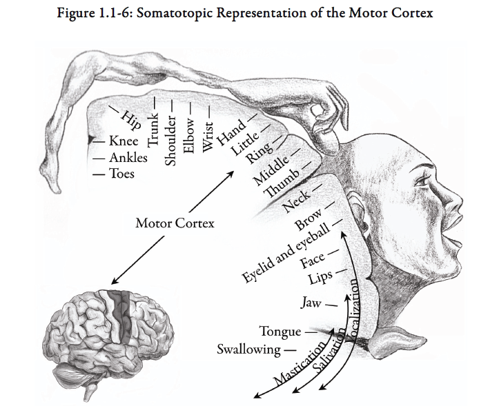
Surrounding the central sulcus are two long gyri that are important in initiating movement and in interpreting sensations from the body. Generally, these gyri are distinguished from each other such that the frontal lobe gyrus is called the motor cortex* and the parietal lobe gyrus is called the somesthetic cortex* (because it controls external body sensations). These terminologies are used to emphasize the differences between these cortical areas. In fact, these areas work closely together and are often referred to as the sensory‑motor cortex* to stress their interdependence.
The body is represented somatotopically on this portion of the cortex. Somatotopic representation means that each body surface location is represented on a specific segment of the cortex (See Figure 1.1-6).
-
-
- Association areas
-
Anterior to the motor cortex are areas involved in organizing motor behaviours. One of the motor association areas is involved in speech, another is critical for eye movements, a third in organizing complex sequences of movement. The motor cortex is primarily involved in executing voluntary movements. Localized stimulation of the motor cortex results in limb or body movements as defined by the somatotopic map (Figure 1.1-6). Lesions* of the motor cortex produce remarkably little deficit in control of most body muscles. The greatest loss is reduced control of speech articulation and fine voluntary movements of the fingers.
Posterior to the sensory‑motor cortex are association areas for body sense. Damage to one of these areas (especially in the right cerebral hemisphere) leads to the feeling that parts of the body (e.g., the legs) are not your own, but are simply foreign objects attached to your body.
In summary, the cerebrum is the largest part of the brain. It is responsible for consciousness and governs intelligence and reason. Although much is known about the functions of the rest of the cerebral cortex, there are still large areas whose functions are not well defined.
Reticular Formation
Scattered throughout the pons and medulla and extending up into the midbrain are numerous large and small neurons that constitute the reticular formation or the reticular activating system (RAS)*. These are considered to be the brain’s “watchdog”, operating during periods of wakefulness to keep the mind awake and alert. If impulses from the RAS are diminished the individual will fall asleep. Damage to these structures may result in unconsciousness or prolonged coma*.
Limbic System
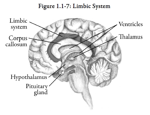
The limbic system* is a collection of structures which roughly form a ring around the corpus callosum (see Figure 1.1-7).
The function of the limbic system is related to the expression of emotion so it is sometimes referred to as the “emotional brain”. The system functions in a way which is not yet understood to make us experience a range of emotions including fear, anger, sorrow, pleasure and sexual feelings.
Cerebrospinal Fluid
Within the cerebrum lie cavities, called ventricles, where cerebrospinal fluid (CSF)* is produced.
This fluid bathes the brain tissue and carries away metabolic waste as it drains down the spinal cord. The CSF also acts as a shock absorber if there is a shift in body or head position. It also maintains homeostasis in the CNS.
Thalamus and Hypothalamus
The thalamus* and the hypothalamus* are located at the upper end of the brainstem (see Figure 1.1-7).
The thalamus is a centre for routing information to the cerebral hemispheres. For example, sensory systems (such as the ones that transmit pain and touch) have a relay centre in the thalamus on the way to the cerebral cortex. The hypothalamus, cerebellum, and brainstem also relay information through the thalamus. Therefore, the functions represented by the thalamus include motor functions and integrative (e.g., sensory‑motor interaction) functions.
The hypothalamus is a collection of neurons located just below the thalamus. The hypothalamus coordinates autonomic activity and serves as a relay station between the cerebral cortex and the lower autonomic and spinal cord somatic centres. The hypothalamus is involved in co-ordinating such behaviours as sleep, appetite, emotions and temperature regulation. The hypothalamus also regulates endocrine function, since it controls the activity of the nearby pituitary gland by secretion of releasing hormones.
Basal Ganglia
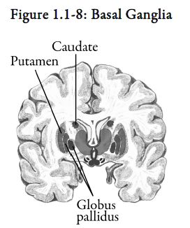
Lying underneath the cerebral cortex is a large system called the basal ganglia*. The structures of the basal ganglia are large grey matter regions that have connections from the brainstem and cerebral cortex. Three principal components of the basal ganglia include the following:
-
-
- neostriatum* (composed of the caudate nucleus*, putamen* and nucleus accumbens)
- globus pallidus*
- substantia nigra
-
These three principal components (see Figure 1.1-8) form the core of the extrapyramidal motor system (EPS)*.
The basal ganglia has an important role in the propagation of movement. The EPS is responsible for maintenance of posture, muscle tone and the modulation of voluntary movement.
Meninges
The meninges* consist of three connective tissue membranes that cover and protect the brain and spinal cord:
-
-
- dura mater* — tough, outermost layer
- arachnoid* — the web-like, middle layer
- pia mater* — innermost, protective layer
-
The space between the dura mater and the arachnoid is called the subdural space*. The subarachnoid space* is the space between the arachnoid mater and the pia mater.
Blood-Brain Barrier
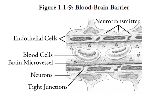
The walls of the brain capillaries are specialized in that they are relatively impermeable compared to other capillaries in the body. This limits the movement of substances from the blood into the extracellular fluid of the brain. Only water, glucose, some amino acids and respiratory gases pass readily through these specialized capillary walls. This protects the delicate neuronal tissue of the brain from toxic insults and prevents the activation of uncontrolled neuronal activity. During CNS infections, the blood-brain barrier is less resistant to penetration of drugs.
The ability of a drug to cross the blood-brain barrier is a key therapeutic consideration when treating disorders of the CNS. Lipid soluble (nonionized) drug molecules, not bound to plasma proteins, can diffuse easily through the blood-brain barrier and enter the brain.
Summary — Section 1: Anatomical and Functional Organization of the Brain
The central nervous system (CNS) is composed of the spinal cord and brain. The function of the CNS is to receive sensory information from organs and receptors in the body, analyze the information and initiate a response.
The brain controls thought, movement and many body processes. The main structures in the brain are the brainstem, cerebellum, and the cerebrum. The brainstem provides a pathway for messages from the brain to the body and the body to the brain, and connects with 10 of the 12 pairs of cranial nerves that control basic functions such as breathing, blood pressure, and eye reflexes. The brainstem consists of the medulla, pons, and midbrain. The cerebellum plays a major role in co-ordinating body movement. The cerebrum consists of the right and left cerebral hemispheres connected at the corpus callosum. Each cerebral hemisphere contains four lobes – frontal lobe, parietal lobe, occipital lobe and temporal lobe. The outer layer of the cerebrum, called the cerebral cortex, contains specialized functional areas – the visual cortex, auditory cortex, sensory-motor cortex, and association areas.
The reticular activating system (RAS) is the brain’s “watchdog”, operating during periods of wakefulness to keep the mind awake and alert.
The limbic system is related to the expression of emotion so it is sometimes referred to as the “emotional brain”.
Cerebrospinal fluid, contained within cavities of the cerebrum called ventricles, bathes the brain tissue and carries away metabolic waste as it drains down the spinal cord. The CSF also acts as a shock absorber if there is a shift in body or head position and it maintains homeostasis of the CNS.
The thalamus routes information to the cerebral hemispheres. The hypothalamus, located just below the thalamus, coordinates autonomic activity such as sleep, appetite, emotions, and temperature regulation.
The basal ganglia located underneath the cerebral cortex contains structures that play an important role in maintaining posture, muscle tone and ordered voluntary movement.
The CNS is covered by the meninges – the dura mater, arachnoid membrane, and the pia mater.
The brain capillaries are relatively impermeable compared to other capillaries in the body. This blood-brain barrier limits the movement of substances from the blood into the extracellular fluid of the brain. The barrier is less efficient during times of CNS infection or inflammation.
Progress Check — Section 1: Anatomical and Functional Organization of the Brain
1.
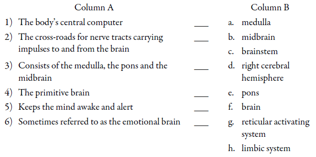
Match the description in Column A with the corresponding anatomical term in Column B. Terms from Column B may be used more than once, or not at all.
2.
The last upper inch of the brainstem is called the _______________________.
3.
The _______________________ fissure divides the cerebrum into the right and left cerebral hemispheres.
4.
Name the four major lobes of the brain.
1) _______________________________
2) _______________________________
3) _______________________________
4) _______________________________
5.
The mass of nerve fibres that connects the two cerebral hemispheres is called _________________________.
6.
Sulci are _______________________.
Gyri are _______________________.
7.
Indicate whether each of the following statements is true or false.

8.
Why are artists often described as being “right-brained” and engineers as being “left-brained”?
______________________________
______________________________
______________________________
9.
What is the function of the extrapyramidal motor system?
______________________________
______________________________
______________________________
10.
The frontal lobe in humans is believed to be important for:
______________________________
______________________________
11.
The three meningeal layers that cover and protect the CNS are called:
1) _____________________________
2) _____________________________
3) _____________________________
12.
The purpose of the blood-brain barrier is to:
______________________________
______________________________
Progress Check Answers — Section 1: Anatomical and Functional Organization of the Brain
1.
1) f. brain
2) a. medulla
3) c. brainstem
4) c. brainstem
5) g. reticular activating system
6) h. limbic system
2.
midbrain
3.
medial longitudinal
4.
1) frontal lobe
2) parietal lobe
3) temporal lobe
4) occipital lobe
5.
the corpsus callosum
6.
small fissures
bumps between sulci
7.
1) False – The visual cortex is located at the posterior of the brain.
2) True
3) True
8.
The right side of the cerebrum processes pattern and space perception, while the left side processes symbols and logic.
9.
The EPS maintains posture and muscle tone, and modulates voluntary movement.
10.
long term plans and complex moral judgments
11.
1) the dura mater
2) the arachnoid
3) the pia mater
12.
protect the delicate neuronal tissue of the brain from toxic insults
Section 2 – Neurotransmission
Learning Objectives
After reading this section, you should be able to:
- State the function of the structural components of a neuron.
- Describe how impulses are conducted within the nervous system.
- Describe how neurotransmitters interact with receptors to produce a response.
- Describe how a nerve impulse is terminated.
- Identify the major neurotransmitters that facilitate neuronal transmission in the brain.
- Explain how dysfunction of specific neurotransmitter systems might relate to psychiatric disorders.
Neurons
The human brain is comprised of billions of nervous system cells known as neurons*. Each neuron is a microscopic information processing system that receives and transmits messages to and from other cells.
Neurons have a nucleus, a membrane, and a variety of structures within the cell body, just as other cells do. However, neurons, unlike many other cells, do not possess the ability to regenerate under normal circumstances. This explains why damage to nerve structures may lead to permanent sequelae. Also, neurons do not look like other cells because they have dendrites* and axons* extending from the body of the cell. (See Figure 1.2-1)
Dendrites are located at the receptive end of the neuron and conduct impulses toward the body of the nerve cell. Depending on the type of neuron, it may have hundreds of branching dendrites. Within the cell body, the signals received are integrated to produce a single output to the axon.
Axons are nerve fibres that conduct impulses away from cell bodies to the target cells or tissues.
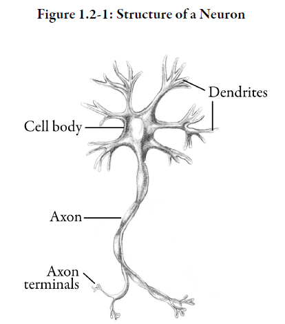
Therefore, neurons are described as having a bipolar structure. Signals are received at one end of the cell and output is transmitted from the other end.
Each neuron has only one axon. However, depending on the location of the neuron, its axon may be very short (< 1 mm) or very long (e.g., extending from the lumbar spine to the toe).
Most axons are coated with a fatty, insulating layer called a myelin sheath*. The presence of myelin increases the speed of transmission of electrical impulses along the axon, and also helps prevent loss of electrical charge.
Neurotransmission
Neurotransmission refers to the transmission of an electrical impulse along the membrane of a neuron and the chemical transmission of messages between nerve cells and target cells or tissues. The impulse travels along the membrane of the axon. This membrane lets in some charged particles (ions) and keeps others out.
1) Resting state (polarized)
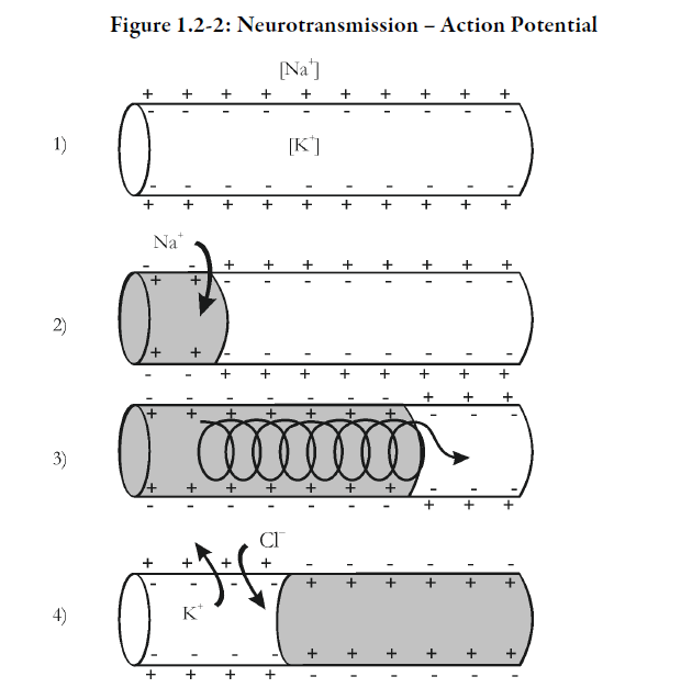
In the resting state (i.e., the neuron is not being stimulated), the inside of the axon is negatively charged with respect to the outside, and the membrane is said to be polarized. The major extracellular ion is sodium (Na+); the major intracellular ion is potassium (K+). See Figure 1.2‑2, #1.
2) Action potential (depolarized)
When a part of the axon is stimulated, the permeability of the membrane is changed, allowing sodium ions to flood into the axon. This causes a local reversal of charge and the cell is said to be depolarized. The electrical impulse, also referred to as an action potential*, has begun. See Figure 1.2-2, #2.
The action potential in one area of the axon acts as a stimulus for another action potential in the next area of the axon and the process repeats itself. This causes the action potential to move down the axon in the form of an electrochemical wave. See Figure 1.2-2, #3.
The wave travels in one direction since a new action potential can only be stimulated in an area of the axon that is in its resting state (or polarized).
Action potentials only occur when the net movement of positive charge through the ion channels is inward. The point at which this occurs is known as the threshold. As long as the stimulus is strong enough to exceed this threshold, the action potential will occur. Stronger stimuli will not result in larger action potentials. This property of action potentials occurring maximally or not occurring at all is sometimes referred to as an all-or-none principle.
3) Repolarized
Following depolarization, negatively charged chloride ions pass into the axon and positively charged potassium ions pass out of the axon. This causes the inside of the axon to become negatively charged once again and the cell is said to be repolarized. See Figure 1.2-2, #4.
Finally, the sodium-potassium pump, using cellular energy in the form of ATP* (adenosine triphosphate), pumps excess sodium ions out of the cell and potassium ions back into the cell. This restores the initial concentration of sodium and potassium ions inside and outside of the nerve cell.
Synapse
Each neuron is a separate cell. Terminals at the end of the axon link a neuron to other neurons. The nerve terminals have a characteristic bead-like appearance. The space between the terminal on the axon of one neuron and the dendrites of another is known as a synapse* (or synaptic cleft). A presynaptic neuron conducts an impulse toward a synapse. An impulse is conducted away from a synapse by a postsynaptic neuron. See Figure 1.2-3.
A typical brain cell has between one thousand and ten thousand synapses. Each synapse acts as a tiny electrochemical crossroad for the movement of electrical impulses.
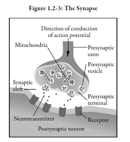
Vesicles located in terminals in presynaptic neurons are filled with neurotransmitters*, which are specialized chemical messengers released in response to nerve stimulation.
When the action potential moving along the axon reaches the terminal of the presynaptic axon, it causes the presynaptic vesicles to burst open and release neurotransmitters into the synaptic cleft. The neurotransmitter is detected by receptors on the dendrite of a receiving cell (a postsynaptic cell) and an electrochemical wave is initiated in that neuron. This process of signal passage from presynaptic to postsynaptic cells continues until the electrical impulse reaches its destination. In summary, electrical impulses are propagated from one neuron to another by the release of messenger chemicals or neurotransmitters into the synapse.
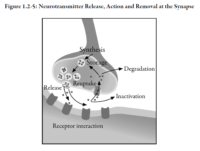
After interacting with the receptor on the receiving cell, the neurotransmitter is either destroyed by enzymes, or there is reuptake of the neurotransmitter by the presynaptic vesicles and the neurotransmitter is stored for further use.
Not every receptor contacted by neurotransmitters passes on the electrical message. Many synapses are in fact inhibitory and prevent the receiving cell from firing. A single neuron may have many excitatory and inhibitory synapses and a constant interplay between these receptor types determines whether a particular neuron fires (passes on the impulse) or not.
In addition to postsynaptic receptors, there are also presynaptic receptors on the terminal buttons of some neurons. These presynaptic receptors monitor the concentration of neurotransmitter within the synaptic cleft, providing feedback that controls the further synthesis, release and degradation of neurotransmitters. They are referred to as autoreceptors*.
This complex electrochemical process, occurring in billions of neurons, regulates the functions of the nervous system. Unfortunately, it may become dysfunctional through a number of mechanisms. For example, if the amount of calcium or potassium in the synaptic region (the electrolytes* that are involved in action potential propagation and stimulation of vesicle release) changes slightly, then fewer vesicles will be activated and transmission may not occur.
If neurotransmitter abnormalities occur, they may result in neurological sequelae such as seizures* or abnormal movement disorders. For example, Alzheimer’s disease is associated with a deficiency of acetylcholine (ACh)* and Parkinson’s disease* is associated with a deficiency of dopamine*. Abnormalities in neurotransmitter systems also contribute to psychiatric disorders such as depression*, disturbances of thought processing, mood or behaviour.
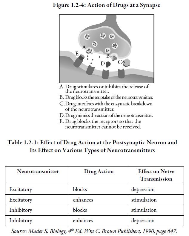
Most nervous system drugs act at the level of the synapse by modifying the levels of neurotransmitters involved in information transfer between neurons. See Figure 1.2-4 and Table 1.2-1 for the action of drugs at a synapse. Generally, only one of these actions occurs at a particular synapse.
Classes of Neurotransmitters
Some neurotransmitters act as both central and peripheral nervous system transmitters. The existence of alpha, beta and acetylcholine receptors in the CNS is a major factor in the use of drugs, particularly in psychiatric practice.
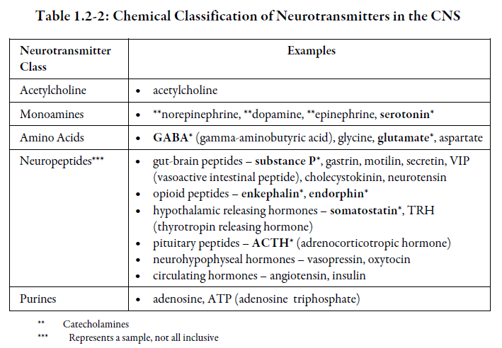
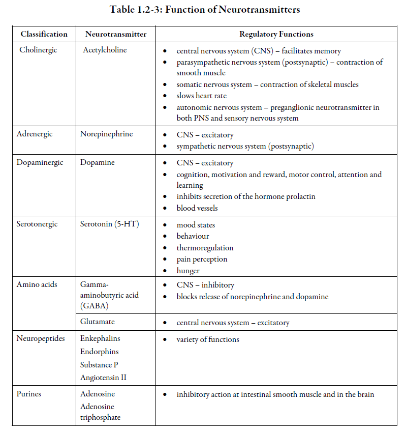
Neurotransmitters can be grouped according to their chemical structure, as shown in the following table.
Did you know?
As many as 100 or more different substances act as neurotransmitters. However, only a little more than 20 have been well studied with respect to their actions. All drugs in psychiatry influence neurotransmitter systems. The rationale for the use of certain drugs and the potential side effects of these drugs can be best understood when the various neurotransmitter systems are taken into consideration.
Each nerve cell releases only one type of transmitter substance, although some recent evidence has suggested that in some cases one or more transmitters may be released together from the same neuron. Neurotransmitters produce their effects by interacting with receptors on the surface of the receiving cell. The shape and electrical charge of the transmitter and its receptor are felt to form the basis for the specificity of the transmitter-receptor interaction.
Each neurotransmitter tends to exert a characteristic action – excitatory or inhibitory, quick or slow, short or long lasting – but sometimes a neurotransmitter may behave differently at different sites. For example, acetylcholine exerts excitatory effects in the brain but in some portions of the peripheral nervous system, such as the heart, it has inhibitory effects.
As previously mentioned, once the neurotransmitter has exerted its effect the action of the substance is terminated by degradation or re-uptake of the neurotransmitter. See Figure 1.2-5.
Although each neuron is generally assumed to release only one neurotransmitter, a single neuron may possess receptors for a variety of different neurotransmitters. This enables a neuron to receive impulses from many other neurons, each capable of exerting a different effect on the receiving neuron.
Abnormalities are known to occur in which specific neurotransmitters are:
-
-
- not synthesized at all
- synthesized in insufficient amounts
- synthesized in excessive amounts
- synthesized but not released properly from synaptic vesicles
- not able to produce the desired receptor effect due to decreased numbers of receptors or receptor insensitivity
- not degraded or taken back up into the synaptic vessels at a normal rate; i.e., too fast or too slow, or degraded by enzymes before they can exert an effect on a receptor
-
These abnormalities may be important contributors to certain psychiatric disorders. The neurotransmitters that have been theorized to play an important role in mental illness are:
-
-
- acetylcholine
- norepinephrine
- dopamine
- serotonin
- gamma-aminobutyric acid (GABA)
- glutamate
-
Acetylcholine (ACh)
Acetylcholine (ACh) is synthesized from choline and acetyl coenzyme A by the enzyme choline acetyltransferase. It is broken down by the enzyme acetylcholinesterase*.
ACh receptors are of two types: muscarinic and nicotinic. In the peripheral nervous system, muscarinic receptors are primarily found in smooth muscle of various organs and may be either excitatory or inhibitory. Atropine antagonizes the effects of ACh at muscarinic receptors.
Nicotinic receptors in the peripheral nervous system are notable for their involvement with contraction of skeletal muscle. Nicotinic receptors are excitatory and can be antagonized with drugs such as succinylcholine, a muscle relaxant.
Acetylcholine is also the main transmitter for the parasympathetic system, and has important effects in the brain. In the cortex, the receptors appear to be predominantly muscarinic. A mixture of nicotinic and muscarinic receptors is found in the thalamus and lower parts of the brain.
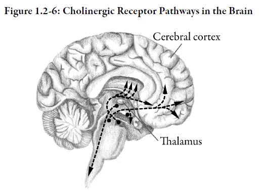
Acetylcholine systems have been linked with arousal mechanisms, temperature regulation and memory. In high doses, atropine (an anticholinergic agent) produces excitation and manic-like effects. This suggests that ACh, through muscarinic receptors, may play a role in mood regulation.
Norepinephrine (NE)
Norepinephrine (NE) is derived from the amino acid phenylalanine through a number of intermediary compounds. Its immediate precursor is dopamine.
The action of norepinephrine is terminated largely by the enzymes catechol-O-methyl transferase (COMT)* and monoamine oxidase (MAO)*. A norepinephrine re-uptake pump also terminates its action by removing it from the synapse and transporting it back into the presynaptic neuron.
There are multiple receptors for norepinephrine. The subtypes can be classified into alpha and beta-receptors. Alpha- and beta-receptors are further subdivided into alpha-1, alpha-2, beta-1 and beta-2 receptors. In addition, different subtypes of these receptors can be found on the presynaptic neuron as well as the postsynaptic neuron. The presynaptic alpha-2 receptor serves as an auto receptor that will turn off further release of norepinephrine when norepinephrine binds to it.
Like acetylcholine, norepinephrine is a neurotransmitter with an important role in the central and peripheral nervous systems. Through stimulation of alpha- and beta-receptors, the sympathetic nervous system plays an important role in regulating the cardiovascular, respiratory and other systems.
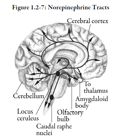
In the brain there are two principal norepinephrine tracts. Those terminating in the hypothalamus and limbic system appear to have a functional role in affective (mood) disorders such as depression and mania*. Those terminating in the cerebral cortex and cerebellum appear to be associated with regulation of the sleep-wake cycle, anxiety*, learning, memory and pain perception.
Dopamine (DA)
In dopaminergic neurons, the amino acid tyrosine is converted to L-DOPA* which is in turn converted to dopamine by a decarboxylating enzyme.
The action of dopamine is terminated by re-uptake or metabolism by the enzymes MAO and COMT.
The effect produced by dopamine is dependent upon the type of receptor, location of receptor (presynaptic vs. postsynaptic) as well as the location of dopaminergic pathway where it exerts its effects. The location of the dopaminergic pathway determines the region of the brain that dopamine affects.
Several types of dopamine receptors have been identified. They regulate dopamine neurotransmission. They may be found on the presynaptic neuron where they act as an autoreceptor or may be found postsynaptically. There are five subtypes of dopamine receptors: D1, D2, D3, D4 and D5. First generation (typical) antipsychotics affect D1 and D2 to produce antipsychotic effects as well as adverse movement disorder effects.
The second generation (atypical) antipsychotics are thought to affect D2 receptors as well, but specifically in the mesolimbic pathway. They also affect other dopamine receptors (D1, D3, D4), but the significance of this is unknown. Second generation antipsychotics also affect serotonin type-2 receptors (5-HT2), and it is thought the interplay between the dopamine and serotonin pathways is responsible in part for their “atypicality”.
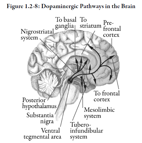
In three of the four major dopamine neuronal pathways, the neuronal cell bodies are contained in the midbrain (Figure 1.2-8). Dopamine neuronal pathways include:
-
-
- the nigrostriatal pathway
- the mesolimbic pathway
- the mesocortical pathway
- the tuberoinfundibular pathway
-
1) Nigrostriatal pathway
The nigrostriatal pathway appears to play a major role in motor coordination. In Parkinson’s disease, the deterioration in motor function appears to be due to a reduction in the number of dopaminergic neurons in this area of the brain. Drugs that upset the dopaminergic system in this region of the brain, such as older, conventional antipsychotics used in the treatment of schizophrenia, can also produce symptoms that mimic Parkinsonism.
2) Mesolimbic pathway
Overactivity of the mesolimbic dopamine pathway, also arising from the midbrain, has been suggested as the cause of many of the signs and symptoms of schizophrenia*. Antipsychotic agents that block this pathway may treat psychotic symptoms seen in schizophrenia.
3) Mesocortical pathway
The mesocortical dopamine pathway may mediate positive and negative symptoms of psychosis*. Some theories suggest that typical antipsychotic agents may mimic or increase the negative symptoms of schizophrenia when blocking this pathway.
4) Tuberoinfundibular pathway
The fourth important dopamine pathway originates from the hypothalamus. The tuberoinfundibular dopamine pathway is involved in the hypothalamic control of the pituitary gland. Dysfunction of this system alters the release of many endocrine substances, such as prolactin* and melanocyte* stimulating hormone. Drugs that disrupt this dopaminergic system, such as older, conventional antipsychotics used to treat schizophrenia, can have endocrine side effects.
Serotonin (5HT Hydroxy-Tryptamine)
Serotonin (5-HT) is synthesized from the amino acid tryptophan by tryptophan hydroxylase* and aromatic amino acid decarboxylase. It is enzymatically degraded by MAO. Serotonin undergoes re-uptake into the presynaptic nerve terminal by a selective serotonin transport pump similar to re-uptake pumps found for norepinephrine and dopamine.
Many subtypes of serotonin receptors have been identified. The most important subtype is 5-HT2A, which is found on the postsynaptic terminal. This receptor is involved in the mechanism of the development of depression. Other postsynaptic subtypes are 5-HT1A, 5-HT1D, 5-HT2C, 5-HT3, and 5-HT4. Presynaptic receptors 5-HT1A and 5-HT1D are autoreceptors which, when serotonin binds to them, decreases further release of serotonin and thus neuronal impulse flow.
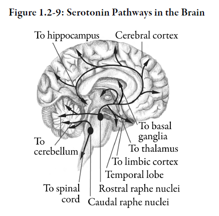
Serotonin neurons in the brain are relatively few in number. They appear to be of major importance in the regulation of mood states, behaviour, thermoregulation, and pain perception. The serotonergic tracts arise in the midbrain and pons and terminate in the cerebellum, cerebral cortex, hypothalamic and pituitary regions (Figure 1.2-9).
Gamma-Aminobutyric Acid (GABA)
GABA is produced from the amino acid glutamate by the enzyme glutamic acid decarboxylase. Its action is terminated by re-uptake or by enzymatic degradation by monoamine oxidase (MAO).
Receptors for GABA are divided into subtypes GABA-A and GABA‑B. GABA-A receptors are gate-keepers for a chloride channel and can be modulated by benzodiazepine receptors.
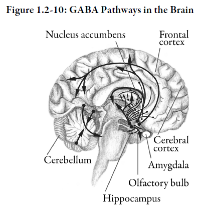
GABA is the most common inhibitory transmitter in the brain and overall may account for transmission in 25% to 40% of all brain synapses (Figure 1.2-10). GABA plays an important role in the regulation of dopaminergic neuronal systems, both by inhibiting dopamine release from synaptic vessels and by modulating dopamine mediated responses postsynaptically. It thus may play an important role in various psychiatric disorders.
Glutamate
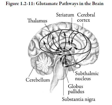
Glutamate is a major excitatory neurotransmitter in the CNS. Excessive activation of glutamatergic synapses causes large influxes of calcium into neurons, which can lead to cell death.
This mechanism of cell death is believed to be important in acute neurological disorders such as stroke and CNS trauma.
Glutamate antagonists are being used therapeutically in the treatment of amyotrophic lateral sclerosis (ALS)* and dementia*.
Neuropeptide Neurotransmitters
Neuropeptides are proteins that consist of larger molecules than norepinephrine and acetylcholine, which are very small molecules.
Some neuropeptides are known to be both hormones and neurotransmitters that act on the brain. For example, the neuropeptides enkephalins, endorphins and Substance P are involved in the perception of pain. Angiotensin II induces a desire to drink when you lose body fluids via the renin-angiotensin-aldosterone pathway that controls sodium and water content in the body.
Purines
Adenosine triphosphate (ATP) is a purine nucleotide. It is released together with ACh, dopamine and norepinephrine. Adenosine triphosphate has inhibitory action at intestinal smooth muscle and possibly in the brain.
Summary — Section 2: Neurotransmission
The neuron is the functional unit of the nervous system. Neurons are composed of a cell body, dendrites, axon, and terminal button.
Neurotransmission refers to the transmission of an electrical impulse along the membrane of a nerve cell, and the chemical transmission of messages between nerve cells and target cells or tissues. A synapse is formed at the junction between the terminal button on the axon of one neuron and the dendrites of another neuron. Synapses can be excitatory synapses or inhibitory synapses.
Electrochemical stimulation at the terminal buttons causes synaptic vesicles to release neurotransmitters into the synaptic cleft. The neurotransmitter reaches a postsynaptic receptor on the dendrite of a receiving cell and initiates either an excitatory or inhibitory process in that receptor. Excitatory actions will perpetuate the stimulus through the receiving neuron. Inhibitory actions will prevent the nervous impulse from moving through the next neuron.
After interacting with the receptor on the receiving cell, the neurotransmitter is either destroyed by enzymes or recaptured by the synaptic vesicles and stored for further use.
Presynaptic receptors (autoreceptors) on the terminal buttons of some neurons monitor the concentration of neurotransmitter within the synaptic cleft. Autoreceptors provide feedback, which controls the further synthesis, release and degradation of neurotransmitters.
Neurotransmitters are subdivided chemically into five classes:
-
-
- acetylcholine
- monoamines – norepinephrine, dopamine, serotonin
- amino acids – GABA, glutamate
- neuropeptides
- purines
-
Although each neuron usually releases only one neurotransmitter, a single neuron may possess receptors for several neurotransmitters. The action of the neurotransmitter is terminated by degradation or re-uptake from the synapse.
Psychiatric disorders may arise if neurotransmitters:
-
-
- are not synthesized at all
- synthesized in insufficient amounts
- synthesized in excessive amounts
- synthesized but not released properly from synaptic vesicles
- not able to produce the desired receptor effect due to decreased numbers of receptors or receptor insensitivity
- not degraded or taken back up into the synaptic vessels at a normal rate
-
Neurotransmitters that play an important role in mental illness are:
-
-
- acetylcholine
- norepinephrine
- dopamine
- serotonin
- gamma-aminobutyric acid (GABA)
- glutamate
-
Acetylcholine is degraded by acetylcholinesterase. Acetylcholine reacts with muscarinic and nicotinic receptors. Peripheral muscarinic receptors (excitatory or inhibitory) are primarily found in smooth muscle. Peripheral nicotinic receptors (excitatory) are involved with contraction of skeletal muscle.
Norepinephrine is degraded by catechol-O-methyl transferase (COMT) and monoamine oxidase (MAO). A re-uptake pump also terminates norepinephrine’s action. Norepinephrine interacts with alpha-1, alpha-2, beta-1 and beta-2 receptors. Presynaptic alpha-2 autoreceptors inhibit release of norepinephrine.
Dopamine’s action is terminated by re-uptake or metabolism by COMT and MAO. Its receptors are called D1, D2, D3, D4 and D5. Dopamine’s effects depend upon the type of receptor, location of receptor (presynaptic vs. postsynaptic) as well as the location of dopaminergic pathway where it exerts its effects. Dopamine neuronal pathways include the nigrostriatal, the mesolimbic, the mesocortical and the tuberoinfundibular pathway. The nigrostriatal pathway plays a major role in motor coordination. Overactivity of the mesolimbic pathway is associated with signs and symptoms of schizophrenia. The mesocortical pathway may mediate positive and negative symptoms of psychosis. The tuberoinfundibular pathway is involved in hypothalamic control of the pituitary gland.
Serotonin is enzymatically degraded by MAO. It undergoes re-uptake into the presynaptic nerve terminal by a selective serotonin transport pump. Serotonin receptors include 5-HT2A (a postsynaptic receptor involved in depression), 5-HT2C, 5-HT3, and 5-HT4. Presynaptic autoreceptors (5-HT1A, 5-HT1D) decrease further release of serotonin. Serotonin receptors are involved in the regulation of mood states, behaviour, body temperature and pain perception.
GABA’s inhibitory action is terminated by re-uptake or by enzymatic degradation by monoamine oxidase. Receptors for GABA are called GABA-A and GABA-B. GABA is thought to help regulate dopaminergic neuronal systems, both by inhibiting dopamine release from synaptic vessels and by modulating dopamine mediated responses postsynaptically.
Other neurotransmitters include glutamate, neuropeptides and purines. Glutamate is a major excitatory neurotransmitter in the CNS. Neuropeptides act on the brain. Adenosine triphosphate is released together with ACh, dopamine and norepinephrine, and may have inhibitory action in the brain.
Progress Check — Section 2: Neurotransmission
1.
Neurons differ from other body cells in that each neuron has:
a. a nucleus in the cell body
b. a cell membrane
c. branches that extend away from the cell body
d. a variety of structures within the cell
2.
Nerve fibres that conduct impulses away from the cell body of a neuron are called _________________________.
3.
Indicate whether each of the following statements is true or false.

4.
Define action potential.
_____________________________________
_____________________________________
5.
List four potential abnormalities that may occur with neurotransmitters.
1) ____________________________________
2) ____________________________________
3) ____________________________________
4) ____________________________________
6.
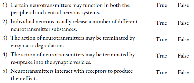
Indicate whether each of the following statements is true or false.
7.
List 4 neurotransmitters commonly involved in mental illness.
1) ____________________________________
2) ____________________________________
3) ____________________________________
4) ____________________________________
8.
Which of the following neurotransmitters is involved in regulating mood and behaviour?
a. glutamate
b. serotonin
c. acetylcholine
d. norepinephrine
9.
Which of the following neurotransmitters is inactivated by MAO? Select all that apply.
a. acetylcholine
b. dopamine
c. GABA
d. glutamate
e. norepinephrine
f. serotonin
Progress Check Answers — Section 2: Neurotransmission
1.
c. branches that extend away from the cell body
2.
axons
3.
1) True
2) False
A synapse may be excitatory or inhibitory.
3) True
4.
An action potential is an electrical impulse that occurs when a neuron sends information down an axon, away from the cell body. An action potential is initiated by depolarization of the nerve cell.
5.
Any 4 of the following:
-
-
- not synthesized at all
- synthesized in insufficient amounts
- synthesized in excessive amounts
- synthesized but not released properly from synaptic vesicles
- not able to produce the desired receptor effect due to decreased numbers of receptors or receptor insensitivity
- not degraded or taken back up into the synaptic vessels at a normal rate
-
6.
1) True
2) False
Each neuron usually releases only one type of neurotransmitter, but some presynaptic neurons can release more than one type.
3) True
4) True
5) True
7.
Any 4 of the following:
-
-
- serotonin
- norepinephrine
- acetylcholine
- GABA
- glutamate
-
8.
b. serotonin
9.
b. dopamine
c. GABA
e. norepinephrine
f. serotonin
Test
1.
The largest structure on the brain is the _________________________.
2.
The sensory-motor cortex in the brain is part of the:
a. thalamus
b. cerebellum
c. cerebrum
d. medulla
3.
Which part of the brain controls conscious mental processes?
a. cerebellum
b. cerebrum
c. pons
d. medulla oblongata
4.
The part of the brain that is sometimes referred to as the “emotional brain” is the _________________________.
5.
The part of the brain that acts as the brain’s “watchdog” and keeps it alert is the _________________________.
6.
The axon of a neuron:
a. has a special membrane that lets in some charged particles (ions) and keeps out others
b. is the cell body of the neuron
c. is not involved in the transmission of nerve impulses
d. contains myelin on its inner surface
7.
The conjunction between the dendrite of one neuron and the axon of another is called a ______________________.
8.
The electrical impulse that travels along a neuron is called _____________________.
9.
What cellular structure stores and releases neurotransmitters?
_________________________________________
10.
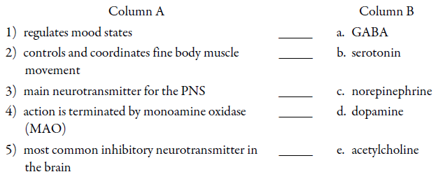
Match the description in Column A with the correct neurotransmitter in Column B. Neurotransmitters in Column B may be selected more than once.
11.
The monitoring of neurotransmitter concentrations in the synaptic cleft is done by:
a. presynaptic autoreceptors
b. postsynaptic receptors
c. oligodendrites
d. mitochondria
12.
Which of the following statements regarding neurotransmitters is true? Select all that apply.
a. The mesocortical dopamine pathway may play a role in schizophrenia.
b. Many drugs can modify the level of neurotransmitters released at the synapse.
c. Norepinephrine and acetylcholine are cholinergic neurotransmitters.
d. Serotonin plays a major role in regulating mood states.
Test Answers
1.
cerebrum
2.
c. cerebrum
3.
b. cerebrum
4.
limbic system
5.
reticular formation
6.
a. has a special membrane that lets in some charged particles (ions) and keeps out others
7.
synapse
8.
an action potential
9.
Neurotransmitters are stored and released from vesicles located in the terminal axon of presynaptic neurons.
10.
1) b. serotonin
2) d. dopamine
3) e. acetylcholine
4) b. serotonin
c. norepinephrine
d. dopamine
5) a. GABA
11.
a. presynaptic autoreceptors
12.
b. Many drugs can modify the level of neurotransmitters released at the synapse.
d. Serotonin plays a major role in regulating mood states.
